Looking for information about other Dental Topics?
Full Website Index• Animated-Teeth.com •

Is a Pre-op X-Ray Always Necessary for a Tooth Extraction?
When are preoperative X-rays needed for an extraction?
When planning for an extraction, one question patients often have is whether taking a pre-op X-ray of their tooth is truly necessary.
While it often seems like just another step and added expense, in reality, an X-ray is an important diagnostic tool that provides your dentist with critical insights into your tooth’s condition, its surrounding bone, and any underlying issues that might complicate its removal.
This guide explains when and why X-rays are needed, the types most frequently taken, and the kinds of information they reveal—ensuring you receive the safest, most effective extraction possible.
Consider this scenario. Will a pre-op X-ray be needed?
- A dentist has a patient in their dental chair who has a tooth that has quite a bit of mobility.
- Related to the tooth’s looseness, it’s pretty clear to everyone that pulling the tooth will be exceedingly easy.
- The patient has no interest in the dentist investigating to see if the tooth can be salvaged.
- They just want it pulled, and as inexpensively as possible.
So, with this type of case, is a pretreatment X-ray really needed for the extraction?
What we’ve tried to describe above is a situation that involves the technically simplest kind of tooth extraction possible.
Not one where explicit knowledge about the tooth’s root(s) anatomy will play a significant role in the act of removing it. (A situation where the benefit of taking an X-ray is clearly obvious.)
Instead, a case where the physics of the extraction process are so overwhelmingly in favor of removal that the patient could have probably just pulled their tooth on their own, if they only had a way to numb it up.
So, even with this type of simple case, is a pretreatment X-ray still always needed?
As you’ve probably already guessed, yes, in most cases you can anticipate that your dentist will still want to take one. The remainder of this page explains the reasons why.
Isn’t taking an X-ray just a way of padding the bill?
Just to get all cynicism out of the way, yes, taking a pretreatment radiograph will add to the cost of your bill. But despite the fact that your dentist may thank their lucky stars that they are in a profession where they can dictate what services are required, in reality, it’s hard to imagine that this reason alone has much to do with the way they practice dentistry. Read on.
What kind of preoperative X-ray is needed to perform a tooth extraction?
1) Characteristics that a pre-op radiograph must have.
Pre and postop periapical X-rays.
- The X-ray image must show the entire tooth, and most certainly 100% of its root portion.
- The tooth’s surrounding bone tissue must be visible so associated pathology and anatomical structures can be evaluated.
- The X-ray’s image quality must the acceptable. (Standards may vary among clinicians. | Non-digital duplicate X-rays are sometimes of poor quality.)
- The dentist must be satisfied with the image’s timeliness (date). Depending on the patient’s current circumstances, X-rays that are months old may still be considered satisfactory.
- Two or more X-rays, taken at different angles, can provide valuable additional information and may be needed.
- For routine (‘simple’) extractions – A small, individual X-ray of the tooth (a ‘PA’ X-ray) is usually all that’s needed.
- If a more-encompassing image has been taken (like a panoramic radiograph), its use is likely suitable for both single and multi-teeth “simple” extraction procedures.
- For more involved extractions, including impacted teeth – A panoramic X-ray, which shows 100% of the patient’s jaws, may be required. Or cone beam (3D) imaging may be required because of the added detail, and therefore information, it can provide.
2) What kind of X-ray is usually taken for an extraction procedure?
a) Periapical X-rays (PA’s).
This is the one- or two-tooth X-ray picture you are probably most familiar with seeing at your dentist’s office.
- The film or digital sensor that’s used to capture the image typically measures on the order of 1 by 1.5 inches.
- A periapical X-ray is meant to capture the whole tooth, including the root. The name itself comes from a term that means “around the tip of the root,” which explains why this type of image shows the tooth from crown to root end.
A periapical (PA) dental radiograph.
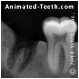
A periapical X-ray should show the entire tooth, including the bone around its roots.
This is the least expensive type of dental X-ray (around $25 to $50). And in the vast majority of cases, this type of picture provides all of the information a dentist needs for a tooth’s extraction procedure.
b) A panoramic film.
This type of radiograph shows all of a patient’s teeth and much of their associated jawbone structure. It’s cost runs on the order of $100 to $130.
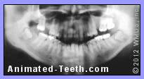
A panoramic dental X-ray.
- A panoramic film excels in revealing information about the relationship between teeth (especially molars) and nearby anatomical structures (such as nerve bundles, sinuses, etc…).
- This has been the standard pre-op imaging technique used with wisdom teeth extraction cases for some decades.
A panoramic view isn’t usually needed for a single routine extraction (with molar extraction being the most likely exception). Although, if it’s expected that the patient will have additional teeth removed in the future, this single X-ray would likely be satisfactory for all of those procedures.
c) Cone Beam CT scan.
Dental cone beam imaging is essentially a CAT scan of your mouth. It produces detailed 3-D views that give your dentist information not available through standard X-rays. However, for most routine extractions, this level of detail isn’t necessary.
Cone beam scans are most often used when planning impacted wisdom tooth extractions, since they allow the dentist to clearly see the tooth’s exact position in relation to nearby anatomical structures (like nerve bundles or sinuses).
▲ Section references – Pogrel, Vandenberghe
3) Which kind of X-ray is usually chosen for an extraction?
a) For routine (“simple”) extractions.
- A periapical film is taken first, with the expectation that it will be satisfactory for the tooth’s extraction procedure.
- Further evaluation, using more extensive/advanced forms of X-ray imaging, is only needed if something that requires greater clarification is noticed on the initial periapical film. (Something that your dentist should be able to show and explain to you.)
b) For multiple extraction cases.
A common exception to taking individual periapical films of individual teeth is when multiple extractions are planned. In this case, it’s common that a panoramic X-ray [or else a full-mouth (complete) series of periapicals] is taken as a cost-saving measure.
c) For impacted wisdom teeth extractions.
A panoramic X-ray is usually taken for impacted wisdom tooth cases because it provides a wide view of the patient’s jawbones. This includes all the wisdom teeth, their roots, and their relationship to nearby anatomical structures like nerves, sinuses, and adjacent teeth. If more detail is needed, then 3-D cone beam imaging is used.
Why Bitewing X-rays aren’t good enough.
It should be mentioned that the routine radiographs that your dentist takes every six months to a year to check for cavities (termed “bitewing” X-rays) are not adequate for performing an extraction. That’s because bitewings don’t show all of each tooth’s root, which of course is the part of most concern to the dentist when performing an extraction.
Taking an X-ray is the prevailing “standard of care.”
For exceedingly easy extractions, like the scenario we described at the top of this page involving a very, very loose tooth, the “standard of care” issue we discuss here probably carries as much weight about why an X-ray is taken as any other factor covered on this page.
The premises of our argument.
- In this country (USA), it’s hard to imagine a dental office that doesn’t have an X-ray machine.
- Even when statistically unlikely, there’s always a chance that important pertinent information might be discovered by taking a pre-op radiograph.
- Taking an X-ray prior to performing an extraction is the way your dentist was trained in dental school.
Now, what if something goes wrong during a patient’s extraction?
If something does go amiss, and no pre-op X-ray was taken, and doing so might have helped to prevent the complication that occurred …
Since essentially all other dentists would have taken a radiograph … (They all have X-ray machines available. Taking a radiograph is the way they were trained in school.) … doing so is considered the prevailing “standard of care.”
So in addition to the distress of having harmed their patient, a dentist could also face serious legal consequences for failing to provide the same level of care that their peers routinely would have.
So, you could say that taking a pre-op X-ray of your tooth helps to ensure that your tooth’s extraction is trouble-free, both for you and your dentist.
There is plenty to learn from taking an X-ray. – Case examples.
Even if a tooth’s removal isn’t expected to pose any challenge, a tooth’s pre-extraction X-ray can still provide the dentist with a lot of beneficial information. And you should want them to have it.
a) It’s important to confirm what problem the tooth has.
Taking an X-ray gives your dentist important additional information about your tooth. And with this information, they can then determine the full range of treatment options they can offer.
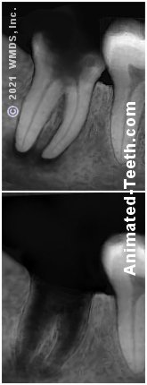
Case example.
The preoperative X-ray shown here reveals a carious lesion (cavity) that has formed in the tooth and shows evidence of nerve death and associated infection.
A severely decayed bicuspid that shows signs of associated infection.

That explains why the tooth hurts. And why, if root canal therapy is not performed, it must be pulled.
This explains why the person has had a history of experiencing throbbing pain. But the patient really has two treatment options that could alleviate that discomfort.
- The X-ray suggests that performing root canal treatment and then rebuilding the tooth would likely provide a successful solution, and a way of saving the tooth.
- Or, if that’s not chosen, the tooth should be extracted.
b) Having additional information is never a bad thing.
Even if everything about the tooth planned for extraction is as expected, an X-ray could reveal other valuable details.
Case example.
Our picture here shows a portion of a panoramic dental X-ray. (By nature, its image is less sharp than periapical radiographs.)
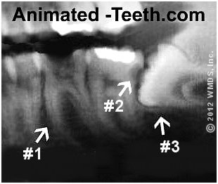
This pre-op X-ray has revealed additional dental problems.
It shows a severely decayed first molar (#1 in our picture) that the patient was already aware of. But the X-ray also shows another, totally unexpected, problem that requires immediate attention too.
The second molar has a significant amount of decay on its distal surface (backside, location #2) where debris has accumulated between it and the impacted wisdom tooth. Most likely the wisdom tooth (#3) will need to be removed before a repair can be made for the 2nd molar.
c) Documentation.
While having an X-ray of a tooth doesn’t necessarily carry more weight than text recorded in a patient’s chart, it certainly can be a valuable aid in documenting (often at just a glance) a tooth’s needed treatment.
- This information might be needed when planning future dental work (like that involving the extracted tooth’s space and/or its adjacent teeth). Also, an X-ray is sometimes requested by insurance companies as a part of determining what benefits will be paid by the patient’s policy.
- With impacted wisdom teeth, taking a pre-op X-ray readily documents the type of impaction involved, which affects billing and insurance issues.
- For more routine extractions, if the added steps of a “surgical” extraction process are performed (another factor that affects the procedure’s cost), a pre-op X-ray can help to document why these steps were needed.
Can’t your dentist just use your old X-rays for your extraction?
Well, in a lot of situations, previously taken X-rays may prove to be perfectly satisfactory. But only the dentist performing the procedure can decide if they are.
Case example.
You had had a full-mouth set of periapical X-rays taken during a previous examination appointment. A problem tooth was identified on them and its treatment was planned. But, somehow, you never followed through and now the tooth has started to cause problems.
If the previous X-rays give evidence of the tooth’s underlying problem, and what the dentist sees now seems to be a logical progression of that condition, they may feel that they have all of the information they require to remove the tooth without taking a new picture.
Some final thoughts.
So, is a pre-extraction X-ray always needed?
In nearly all cases, yes, your dentist will probably insist on one. That’s because they know that even when an extraction looks straightforward, taking an X-ray gives them further insight into the tooth’s roots, surrounding bone, and any hidden problems that could affect the procedure. It’s a safeguard that helps prevent surprises, reduces risks, and ensures the treatment plan is sound.
FYI– If you’re interested in learning more about having teeth removed, we offer pages on these additional topics.
Last reviewed: September 25, 2025
Author: Paul Cotner, DMD — retired dentist.
Published by: WMDS, Inc. — owner of Animated-Teeth.com.
Educational information only — not a substitute for professional dental care.
Page references sources:
Pogrel MA, et al. American Association of Oral and Maxillofacial Surgeons White Paper on Third Molar Data.
Vandenberghe B, et al. Modern dental imaging: a review of the current technology and clinical applications in dental practice. Eur. Radiol. June 2010.